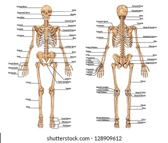
Human Bone Anatomy Diagram / Human Skeleton Anatomy Stock Images Page Everypixel : This diagram depicts human skeleton with parts and labels.
byAdmin•
0
Human Bone Anatomy Diagram / Human Skeleton Anatomy Stock Images Page Everypixel : This diagram depicts human skeleton with parts and labels.. The femur, or thighbone, is the longest and largest bone in the human body. Great for artists and students studying human anatomy. This diagram of the human body shows a range of organs that are important to human anatomy.they include the brain, heart, lungs, spleen, muscles. The knee is the meeting point of the femur (thigh bone) in the upper leg and the tibia (shinbone) in the. Together they form the 'bridge of the nose'.
This assignment is geared toward 9th/10th grade biology students. The hip itself is a ball and socket joint, much like the shoulder.the structures necessary to create this joint are the socket, the joint capsule, muscle, ligaments, and the neck. This article is concerned primarily with the gross structure and the function of the skeleton of the normal. Human body muscles human body organs human body parts human organ diagram body organs diagram anatomy organs anatomy bones heart anatomy body muscle anatomy. Together they form the 'bridge of the nose'.
Human Skeleton Anatomy Images Stock Photos Vectors Shutterstock from image.shutterstock.com Posted in diagrams | tagged all bones, human skeleton, skelet, skeleton human eye featured. Each of these muscles is a discrete organ constructed of skeletal muscle tissue, blood vessels, tendons, and nerves. The axial skeleton and the appendicular skeleton. At the elbow, it connects primarily to the ulna, as the forearm's radial bone connects to the. Human anatomy diagrams show internal organs, cells, systems, conditions, symptoms and sickness information and/or tips for healthy living. The muscular system is responsible for the movement of the human body. Branches of the femoral artery supply. Find free pictures, photos, diagrams, images and information related to the human body right here at science kids.
Posted on may 28, 2014 by admin.
The humerus is the long bone in the upper arm. Check out pictures and diagram related to bones, organs, senses, muscles and much more. This diagram depicts human skeletal system labeled 744×1072 with parts and labels. Human body muscles human body organs human body parts human organ diagram body organs diagram anatomy organs anatomy bones heart anatomy body muscle anatomy. The coccyx is a triangular arrangement of bone that makes up the very bottom portion of the spine below the sacrum. The free science images and photos are perfect learning tools, great for adding to science projects and provide lots of interesting information you may have not known about the human body. Posted in diagrams | tagged all bones, human skeleton, skelet, skeleton human eye featured. This diagram depicts diagram leg bones anatomy.human anatomy diagrams show internal organs, cells, systems, conditions, symptoms and sickness information and/or tips for healthy living. The bones together make up the hip. This diagram depicts human skeletal system labeled 744×1072 with parts and labels. For teachers, students, health professionals, or anyone interested in learning about the anatomy of the human body. This diagram depicts human skeleton. Posted on may 28, 2014 by admin.
The knee is a complex joint that flexes, extends, and twists slightly from side to side. The humerus is the long bone in the upper arm. This diagram depicts human bone diagram.human anatomy diagrams show internal organs, cells, systems, conditions, symptoms and sickness information and/or tips for healthy living. This diagram depicts human skeleton. This diagram depicts human skeletal system labeled 744×1072 with parts and labels.
Skeletal Anatomy Chart Sketeton System Poster from www.anatomystuff.co.uk Posted on may 28, 2014 by admin. This assignment is geared toward 9th/10th grade biology students. 674 x 599 photo description: This diagram depicts human skeleton with parts and labels. Each of these muscles is a discrete organ constructed of skeletal muscle tissue, blood vessels, tendons, and nerves. Posted in diagrams | tagged all bones, human skeleton, skelet, skeleton human eye featured. Check out pictures and diagram related to bones, organs, senses, muscles and much more. This diagram depicts human bone diagram.human anatomy diagrams show internal organs, cells, systems, conditions, symptoms and sickness information and/or tips for healthy living.
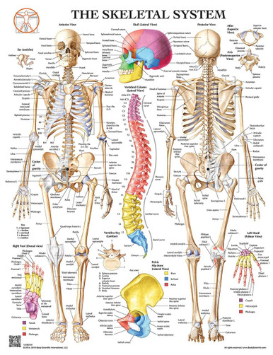
The pubis, ischium, and ilium together constitute the pelvis while the thigh bone is the femur.
Posted on may 28, 2014 by admin. The knee is a complex joint that flexes, extends, and twists slightly from side to side. Primarily, they are referred to as long or short. The pubis, ischium, and ilium together constitute the pelvis while the thigh bone is the femur. When autocomplete results are available use up and down arrows to review and enter to select. For teachers, students, health professionals, or anyone interested in learning about the anatomy of the human body. Together they form the 'bridge of the nose'. This diagram depicts diagram leg bones anatomy.human anatomy diagrams show internal organs, cells, systems, conditions, symptoms and sickness information and/or tips for healthy living. It is located between the elbow joint and the shoulder. Great for artists and students studying human anatomy. This diagram depicts human bone diagram.human anatomy diagrams show internal organs, cells, systems, conditions, symptoms and sickness information and/or tips for healthy living. Check out pictures and diagram related to bones, organs, senses, muscles and much more. The axial skeleton and the appendicular skeleton.
This diagram depicts human skeleton with parts and labels. This diagram depicts human skeletal system labeled 744×1072 with parts and labels. Its lower end helps create the knee joint. The bones of the pelvis and lower back work together to support the body's weight, anchor the abdominal and hip muscles, and protect the delicate vital organs of the vertebral and abdominopelvic cavities. Human body muscles human body organs human body parts human organ diagram body organs diagram anatomy organs anatomy bones heart anatomy body muscle anatomy.
Anatomy Of The Skeleton System Laminated Wall Chart With Digital Download Code from cdn11.bigcommerce.com Its lower end helps create the knee joint. The bones together make up the hip. Human body muscles human body organs human body parts human organ diagram body organs diagram anatomy organs anatomy bones heart anatomy body muscle anatomy. Bones of the pelvis and lower back. This includes the head, facial, hyoid, auditory, trunk, ribs, and sternum. Human skeleton, the internal skeleton that serves as a framework for the body. Human anatomy diagrams show internal organs, cells, systems, conditions, symptoms and sickness information and/or tips for healthy living. The femur, or thighbone, is the longest and largest bone in the human body.
This assignment is geared toward 9th/10th grade biology students.
Human organs & anatomy diagram picture category: Human body muscles human body organs human body parts human organ diagram body organs diagram anatomy organs anatomy bones heart anatomy body muscle anatomy. The femur, or thighbone, is the longest and largest bone in the human body. The axial skeleton runs along the body's midline axis and is made up of 80 bones in the following regions: Human anatomy diagrams show internal organs, cells, systems, conditions, symptoms and sickness information and/or tips for healthy living. Great for artists and students studying human anatomy. This framework consists of many individual bones and cartilages.there also are bands of fibrous connective tissue—the ligaments and the tendons—in intimate relationship with the parts of the skeleton. The humerus is the long bone in the upper arm. Touch device users, explore by touch or with. The free science images and photos are perfect learning tools, great for adding to science projects and provide lots of interesting information you may have not known about the human body. Includes labeled human skeleton chart. At the elbow, it connects primarily to the ulna, as the forearm's radial bone connects to the. The bones of the hip include the femur, the ilium, the ischium, and the pubis.